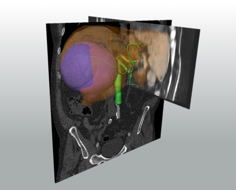
During radioembolization planning, the intervention and the assessment of the therapy, a variety of morphological and functional patient images is acquired: CT and MR image sequences provide anatomical information of the localization of the tumor volume inside the liver. Significant liver parameters, such as liver perfusion or diffusion, can also be derived from these image sequences. Imaging the arterial vessel system inside the liver provides valuable information for the intervention. CBCT and projection images are acquired during the procedure to ensure correct radioembolization. Afterwards SPECT and PET images are used to provide information about the distribution of radionuclides.
Image registration enables the joint use of information from different image modalities and provides a spatial correlation. This is the main reason why image registration plays a central role in radioembolization therapy planning, interventional support and therapy assessment.
The focus in SIRTOP is to provide a smart combination of image registration techniques tailored to liver radioembolization.
 Fraunhofer Institute for Digital Medicine MEVIS